
Science Topics – 112
The cerebral cortex of mammals consists of six stacked layers, each of which has unique combination of cell types and anatomical connections. Although previous studies reported layer-specific neural coding in primary sensory/motor cortices, functional laminar differences of higher association cortex that processes high-level cognition such as memory or attention remain elusive. Here we recorded single-unit responses while monkeys were performing a cued-recall memory task, and reliably localized the position of neurons at laminar resolution using a novel MRI-assisted electrophysiological method. With this method, we mapped laminar distribution of memory-related neurons in area 36 of the temporal cortex and found functional differences of layers during memory recall. Specifically, transformation of neural representations from a cue stimulus to a to-be-recalled target stimulus occurred at the infragranular layers, starting at layer V and then followed by layer VI. While layer V neurons encoded both cue and target information, subset of layer VI neurons showed more exclusive coding of the target stimulus. Neurons in layers II-IV tended to encode cue information rather than target information. These results showed information processing flow from layer V to VI implementing cue-to-target conversion for object memory recall in the primate temporal cortex.
Laminar Module Cascade from Layer 5 to 6 Implementing Cue-to-Target Conversion for Object Memory Retrieval in the Primate Temporal Cortex. Kenji W. Koyano, Masaki Takeda, Teppei Matsui, Toshiyuki Hirabayashi, Yohei Ohashi, Yasushi Miyashita. Neuron 92, 2016. DOI: HYPERLINK "http://dx.doi.org/10.1016/j.neuron.2016.09.024"
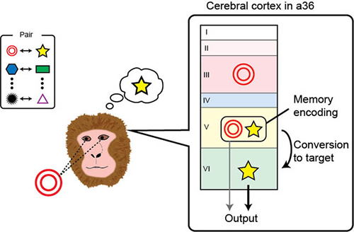
fig. Information processing in monkey area 36 (a36) of the temporal cortex during memory recall. Monkeys were trained to recall target (star) from cue (double circle) stimulus. During information processing from layer V to VI, cue stimulus information is converted into target information. The stimulus images in the figure are modified from the actual one for simplicity.
1 Department of Physiology, The University of Tokyo School of Medicine, Japan
2 Juntendo University Graduate School of Medicine, Japan
(3 Present address: Section on Cognitive Neurophysiology and Imaging, National Institute of Mental Health, USA)