
Science Topics – 126
Voltage-sensing phosphatase (VSP) has a phosphoinositide phosphatase activity regulated by membrane potential change, which consists of two regions, a voltage sensor domain (VSD) similar to that of voltage-gated ion channels and a PTEN-like cytoplasmic catalytic region (CCR). Although the coupling mechanisms between the VSD and the CCR have been studied for a long time, it remains unclear. In this time, we report a novel site on protein-membrane interface (we call “the hydrophobic spine”), which plays a key role in the coupling. Our computer simulations suggest that the nature of the hydrophobic spine influences the membrane binding of the CCR of Ciona intestinalis VSP (Ci-VSP) and its activity. To verify the suggestion, we performed electrophysiological analysis with mutations in the hydrophobic spine of Ci-VSP. These results show that the voltage-dependent phosphatase activity of Ci-VSP depends on the hydrophobicity and presence of an aromatic ring in the hydrophobic spine. To gain more insight, analysis of voltage-dependent conformational changes of VSP using voltage clamp fluorometry (VCF) with fluorescent dye labeled or unnatural amino acid incorporated Ci-VSP. VCF results suggest that VSP has two active states with different enzyme activities and that the nature of the hydrophobic spine affects the latter transition, accompanied by changing the apparent activity of VSP.
Kawanabe A, Hashimoto M, Nishizawa M, Nishizawa K, Narita H, Yonezawa T, Jinno Y, Sakata S, Nakagawa A, Okamura Y.
The hydrophobic nature of a novel membrane interface regulates the enzyme activity of a voltage-sensing phosphatase.
eLife, 7:e41653, 2018.
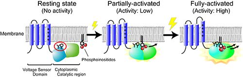
Activation mechanism of voltage sensing phosphatase (VSP): The voltage sensor domain (VSD) is activated by membrane potential change, inducing the conformational changes of the cytoplasmic catalytic region (CCR) and the regulation of its phosphatase activity toward phosphoinositides. A red circle shows “the hydrophobic spine”. Modified from figure 11 of Kawanabe et al. (2018) elife.
Integrative Physiology, Department of Physiology, Graduate School of Medicine, Osaka University, Japan