
Science Topics – 154
The cerebellum contains highest levels of protein kinase C (PKC) in the central nervous system, in which γ isoform of PKC (PKCγ) accounts for more than a half amount of PKC. PKCγ is solely expressed in cerebellar Purkinje cells (PCs). Previous studies using PKCγ-deficient mice exhibiting motor incoordination have shown a critical involvement of PKCγ for climbing fiber (CF) synapse pruning from developing PCs. PKCγ continues to express even after maturation of mice; however, the physiological significance of PKCγ in mature mouse PCs remained unknown.
Using adeno-associated virus vector (AAV), we re-expressed PKCγ in PCs of mature PKCγ-deficient mice. Then, motor incoordination was significantly restored without influencing the multiple CF innervation of PCs. Next, we removed PKCγ from PCs of normally developed adult PKCγ-floxed mice by cerebellar injection of AAV expressing Cre specifically in PCs, leading to emergence of motor defect without affecting a single CF innervation of PCs. These results suggest that PKCγ plays a critical role in motor coordination by a mechanism distinct from CF synapse pruning.
Subsequent experiments revealed that PKCγ suppresses BK channel, a calcium-dependent potassium channel. Depolarization induced by a CF input activates BK channels along the PC dendrites, resulting in electrical shunting. Activated PKCγ suppresses BK channel function, which inhibits declining of CF-triggered electrical activity, resulting in regulation of complex spike waveform and eventually, motor coordination.
Protein kinase Cγ in cerebellar Purkinje cells regulates Ca2+-activated large-conductance K+ channels and motor coordination.
Masashi Watanave, Nobutaka Takahashi, Nobutake Hosoi, Ayumu Konno, Hikaru Yamamoto, Hiroyuki Yasui, Mika Kawachi, Takuro Horii, Yasunori Matsuzaki, Izuho Hatada, Hirokazu Hirai.
Proceedings of the National Academy of Sciences
: 119(7) e2113336119-e2113336119, 2022.
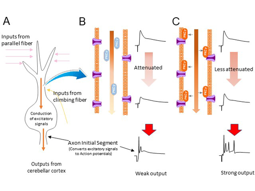
<Figure Legends>
(A) Purkinje cells receive signals from parallel and climbing fibers and transmit them to the axon initial segment where they are converted into a final output signal (complex spike). Complex spikes w and output outside the cerebellar cortex. (B) BK channels are present along the plasma membrane of Purkinje cell dendrites and attenuate the signal traveling down the dendrites. (C) When PKCγ is activated, it translocases to the plasma membrane of dendrites. There, PKCγ phosphorylates BK channels and suppresses BK channel function. As a result, the signal through the dendrite is less attenuated and a strong output signal is formed at the axon initial segment.
1: Department of Physiology, Graduate School of Medicine, Gunma University, Japan
2: Department of Morphological Neuroscience, Graduate School of Medicine, Gifu University, Japan (Current address)