
Science Topics – 157
We have developed a technique for observing the structure and "movement" of a sample in a liquid such as a wet organ with a scanning electron microscope. Since the electron microscope has a high resolution of up to 0.5 nm, it is suitable for small-scale observation. However, since the observation is performed under vacuum, it is necessary to fix the sample to be observed so that the water does not evaporate. Therefore, there is a problem that the conventional electron microscope observation can basically only measure the still image of a fixed sample. In this research, we have developed a new electron microscope technology that can observe the fine structure and movement of living cells as they are. We have created a thin film (DET film: Deformable and Electron Transmissive Film) that can withstand the pressure difference between vacuum and atmospheric pressure and does not break, has excellent electron beam permeability and deformability. By covering the observation sample such as wet organs with this DET film and creating a closed space with the sample holder, we succeeded in observing the fine structure and "movement" of the observation sample immersed in the solution with an electron microscope. By combining image analysis, it is possible to measure "movement".
Real-Time Scanning Electron Microscopy of Unfixed Tissue in Solution using a Deformable and Electron-Transmissive Film., Seine A. Shintani, Seiji Yamaguchi, Hiroaki Takadama, Microscopy: dfac030, 2022..
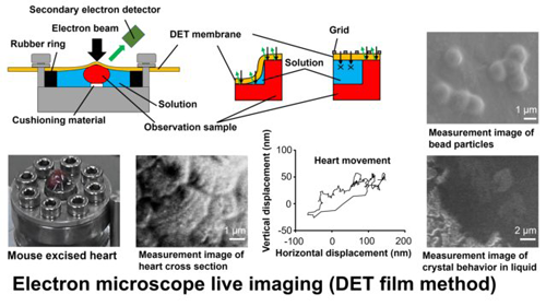
<Figure Legends>
Electron microscope live imaging method (DET film method). Upper left figure: Schematic diagram of the sample holder during observation of the DET film method. Upper right figure: Bead particles observed by the DET film method. Lower left figure: Mouse excised heart set in a sample holder that enables observation of the DET film method. Bottom figure: Cross-sectional image of a mouse-excised heart observed by the DET film method (left) and measurement results of movement (right). Lower right figure: Measurement image of crystal behavior in liquid observed by DET film method.
Department of Biomedical Sciences, College of Life and Health Sciences, Chubu University