
Science Topics – 163
Cellular processes within the cell and across the membrane are mediated by a variety of proteins that sense their respective signals. Voltage-gated ion channels allow ions to pass through the membrane by sensing changes in the membrane potential. Two-pore channels (TPCs) are a member of the voltage-gated ion channel superfamily that responds to a phosphoinositide, PIP2, along with the membrane potential. Each TPC subtype responds to the two types of stimuli differently, however, the detailed mechanisms that account for this diversity have not been fully understood.
We focused on TPC3, a TPC subtype that is primarily activated by membrane potential, and tracked the movement of DII-S4, a center of the voltage sensor, by measuring the fluorescent molecule attached to DII-S4. We found that DII-S4 takes an intermediate state between the "up" and "down" states (see figure). Artificial stabilization of the intermediate state converted TPC3 into a highly PIP2-dependent channel. This result shows that TPCs are a unique type of ion channels that have not only a typical voltage-gating mode achieved by switching between the up and down states of DII-S4, but also have a “hidden mode” corresponding to the intermediate state that is specifically dedicated to the PIP2-gating mode.
Conformational rearrangements in the second voltage sensor domain switch PIP2- and voltage-gating modes in two-pore channels.
Takushi Shimomura, Kiichi Hirazawa, Yoshihiro Kubo.
Proceedings of the National Academy of Sciences of the United States of America
120 (6): e2209569120, 2023.
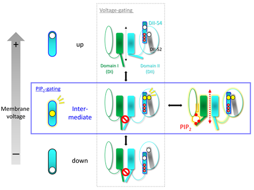
<Figure Legends>
TPC monomer consists of two very similar domains, domain I (DI) and DII. Each domain consists of a pore region (rectangular) that forms an ion-permeation pathway and a voltage sensor (elliptical). The DII-S4 helix in the voltage sensor is the main core for detecting the membrane potential. The (+) on DII-S4 represents a positively charged amino acid residue for detecting the membrane potential. The vertical direction in the figure represents a voltage-dependent mode. In addition, TPC can adopt an intermediate state, in which the binding of PIP2 to DI opens the gate (horizontal). Circles (○) indicate pairs of amino acid residues of DII-S4 and DII-S2 that are in close proximity in the intermediate states, as revealed in this study.
Division of Biophysics and Neurobiology, National Institute for Physiological Sciences, Japan